Sourced by A/Prof Vanessa Panettieri, Editor (Educational), AFOMP Pulse
Over the past six months, the AFOMP Journals—Physical and Engineering Sciences in Medicine, Journal of Medical Physics, and Radiological Physics and Technology—have continued to publish valuable contributions to our field.
Building on insights from the last issue of PULSE, our first focus section delves deeper into AI applications, examining how these advanced tools are transforming auto-segmentation of organs-at-risk structures across various anatomical sites. The featured paper compares commercially available products, offering valuable guidance to help you select the most suitable tool for your clinical practice. We hope it will assist in your current or future evaluations of these technologies.
Our second focus section explores patient treatment safety, highlighting novel approaches in wireless communication for medical implantable devices and the use of a body movement detection system to prevent re-irradiation during radiography. Additionally, we introduce a cost-effective camera system designed for patients undergoing breath-hold irradiation.
In our newly introduced “How to?” section, we examine the impact of mirror systems and flatbed scanner beds on response artifacts in film dosimetry, commonly used in patient-specific quality assurance.
Happy reading! And as always, we welcome your suggestions for topics to explore in future issues.
With contributions kindly provided by Dr Taylah Brennen and Sadia Aftab, Medical Physicists, Peter MacCallum Cancer Centre
1) Focus on: Artificial Intelligence
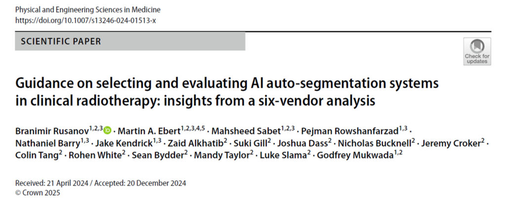
https://doi.org/10.1007/s13246-024-01513-x
This paper by Rusanov et al., published in the Journal of Physical and Engineering Sciences in Medicinepresents a comprehensive evaluation of six vendors’ products providing AI-based auto-segmentation systems, focusing on their performance, integration, data security, vendor support, and ethical considerations. The assessment utilized a subtle set of criteria informed by clinical experience and existing literature, aiming at selecting the safest, least biased, and most efficient systems for clinical use. The authors provide some advice on the process to follow which includes access to trial software which in their view is essential for a thorough assessment of both quantitative and qualitative performance.
In this experience vendors A, B, and D provided trial licenses, while the other thres processed institutional data through secure file transfers. The study identified significant gaps in data security and transparency among all vendors, with most lacking detailed information on data collection, ownership, and de-identification practices. Vendor support tools were generally limited, as most vendors offered limited functionality beyond auto-segmentation, with Vendor B noted for its integration with the Eclipse treatment planning system and additional contour editing tools. Each vendor was scored across seven identified selection criteria, with scores ranging from 1 (poor) to 5 (excellent). Vendor A received the highest weighted score (3.83), indicating better integration and performance, while Vendor E scored the lowest (2.05). The study emphasised the importance of ethical practices in AI applications, including adherence to HIPAA and GDPR regulations. However, there was a general lack of transparency regarding the physical location of cloud servers and data handling. In conclusion, this evaluation provides valuable insights into the current landscape of AI auto-segmentation systems in radiation oncology, emphasizing the need for improved transparency, data security, and vendor support to enhance clinical outcomes. The findings serve as a guide for institutions considering the adoption of such technologies.
2) Focus on: Safety
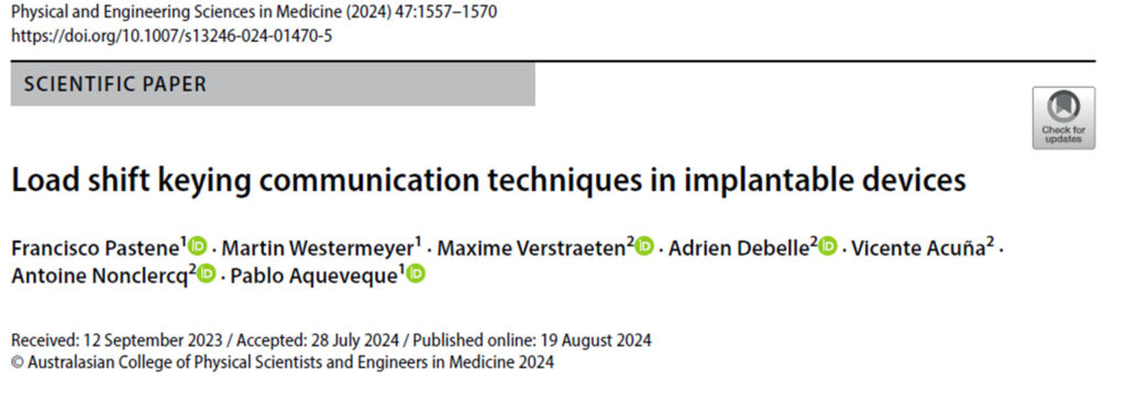
https://doi.org/10.1007/s13246-024-01470-5
This paper by Pastene et al., published in the Journal of Physical and Engineering Sciences in Medicinediscusses advancements in wireless communication for medical implants, specifically focusing on inductive links. Medical implants, used in fields such as orthopedics, cardiac pacemakers, blood pumps, neural implants, and drug delivery systems, perform essential functions by replacing or restoring abilities lost due to diseases or injuries. The study compares three communication techniques: Short-Circuit Technique (SCT), Open-Circuit Technique (OCT), and the newly introduced Disconnected Load Technique (DLT). Each method has distinct operational behaviours. SCT uses a short-circuit across the load for data transmission, OCT disconnects the load, and DLT maintains the load in a disconnected state. This research highlights the importance of stable power delivery to implants and the need for adaptive control strategies to handle variations in coil distance and load conditions. The study also explores the potential for increasing data rates by adjusting carrier frequencies and refining demodulation processes, while ensuring compliance with safety regulations regarding electromagnetic radiation. The methodology involved testing various coil distances and load conditions, focusing on parameters like modulation index and Bit Error Rate (BER). The results indicated that despite using a relatively low carrier frequency of 300 kHz, the techniques achieved adequate data rates for transmitting physiological signals. The findings have significant implications for medical applications, including smart prosthetics, chronic condition monitoring, and drug delivery systems. Although there is no universally accepted standard for communication frequency bands with medical implants, the European Telecommunications Standards Institute (ETSI) suggests the 300-500 kHz range for both powering and communicating with medical implants, which can go up to 31.5 W. The study underscores the necessity of complying with electromagnetic field regulations to ensure safety while developing communication systems for medical implants. The article concludes by acknowledging the study’s limitations, such as the limited number of load scenarios tested, and suggests future research directions to enhance these communication techniques in biomedical engineering, potentially benefiting various stakeholders in healthcare and medical technology.
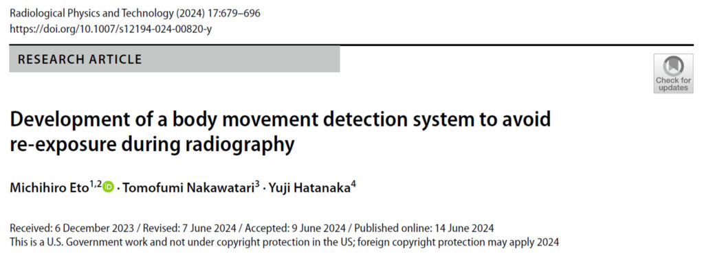
https://doi.org/10.1007/s12194-024-00820-y
This study by Eto et al., published in Radiological Physics and Technology, introduces a body movement detection system designed to prevent unnecessary re-exposure during radiographic examinations of the chest and bones. The system accounts for breathing and involuntary movements, addressing a common challenge in medical imaging for patients who struggle to follow instructions or remain still during scans. Traditionally, post-processing techniques and deep learning tools have been used to correct motion blur after an X-ray is taken. However, this research takes a proactive approach, detecting movement before irradiation occurs. By doing so, it minimizes radiation exposure and improves imaging efficiency in hospitals. The system uses an RGB camera to monitor patients after positioning, analyzing video data in real time using inter-frame difference and optical flow estimation methods. To validate its effectiveness, researchers first tested the system with an anthropomorphic phantom head, simulating body movements using an inclined table and an acrylic plate caster. They then conducted a simulation with three patients, instructing them to assume body positions and breathing patterns similar to those exhibited during PA chest radiography. The results confirmed that the system can accurately detect body wobbles and respiratory motion in real time.
Beyond reducing unnecessary radiation exposure, this tool can also help confirm breath-holding status in patients who have difficulty communicating, ultimately improving radiographic accuracy and patient safety.

https://doi.org/10.4103/jmp.jmp_101_24
As reported in several journals articles commercial systems are now routinely in use in clinical practice in order to account for organs motion and variation, which have been shown to affect delivery of the radiation for areas such as lung, liver or breast. Motion management, as it is often referred to, allows the use of very precise hypofractionation enabling the use of stereotactic treatment for both the brain and the body. Among various technological development the use of sophisticated surface guidance radiotherapy has grown in the last years not only as a tool for patient positioning but also to facilitate the use of breath hold (BH) in certain irradiations. However, some of this technology can be costly limiting its use to only a few departments and often to a limited number of treatment machines. In this work the authors explore the use of a cost-effective Time-of-Flight (ToF) camera system developed to coach patients undergoing treatment in BH, making BH treatments more accessible and reducing treatment delays. Their design is based on the use of a Wi-Fi-compatible Rasberry Pi 3B with and Arducam ToF camera connected in Raspberry Pi case. The interface includes an operator view and a patient view to allow them to observe the breathing pattern. The authors have tested it on volunteers and compared it with a commercial product demonstrating its equivalence and also its feasibility.
3) Focus on: how to?
This section provides practical information on tests and procedures that can be used in the clinic to ensure equipment accuracy and precision.
https://doi.org/10.1007/s13246-024-01478-x
This paper by Shameem et al., published in the Journal of Physical and Engineering Sciences in Medicinefocuses on improving the measurement process of radiochromic film dosimetry, particularly using commercial flatbed scanners, specifically various EPSON models. The research systematically investigates several components of the scanner system including the path length effect and the polarization effects caused by the scanner’s mirror systems.
The aim of this work is to address the orientation effect and lateral response artifacts (LRA) that can affect the accuracy of dosimetry. EBT3 films were prepared and irradiated under controlled conditions, and their responses to varying doses and orientations were analyzed using the ImageJ and Excel software packages, focusing on the red and green color channels. The study examines how the materials used in the scanner bed such as glass and laminating pouches, affect the path length of light and, consequently, the measurement accuracy. It compares materials with different refractive indices to understand their impact on the scanning process. Additionally, the research investigates how the number and quality of mirrors in the scanner system influence the polarization of light, which can affect the readings obtained from the film. The study compares the polarization effects of different scanner models and mirror configurations. The report suggests an optimal scanner configuration that includes avoiding lenses with focal lengths below 50 mm, using glass as the scanner bed material, and limiting the use of mirrors to one or none, with specific angle considerations to minimize LRA effects. Overall, the report contributes to the understanding of how scanner design influences the accuracy of film dosimetry in radiation therapy, providing valuable insights for future improvements in measurement techniques and enhancing the reliability of dosimetry with radiochromic films, which is vital for accurate radiation therapy and patient safety.

