Fujita Health University. Toyoake-city, Japan
Invited Professor
Katsumi Tsujioka
tsujioka@fujita-hu.ac.jp
1. Diagnostic imaging in medical physics
Medical Physics plays an important role in modern medicine. It will be to maintain the health and life of the people, to contribute to the detection and treatment of diseases, and to control radiation exposure. The field of medical physics consists of radiotherapy and diagnostic imaging, but now diagnostic imaging has expanded to include interventional radiology. What about the activities of Medical Physics in such current medical care? In the United State, there is ASTRO (American Society for Radiation Oncology) for Radiation Oncology and AAPM (American Association of Physics in Medicine) for Medical Imaging. In Japan, there is JSMP (Japanese Society of Medical Physics) for Radiation Oncology and JSRT (Japanese Society of Radiological Technology) for Medical Imaging. These are not fully shared. In addition, many members belong to two organizations.
Medical Physics is expected to consist of radiotherapy and diagnostic imaging. And the field of diagnostic imaging has expanded tremendously in recent years.
2. Diagnostic imaging activities in Japan
JRC(Japan Radiology Congress) is made up of three organizations, JSRT, JSMP, and JRS (Japan Radiological Society), and is one of the largest conferences in the world after RSNA (Radiological Society of North America) and ECR (European Congress of Radiology).
JSRT is the main activity society for diagnostic imaging in Japan. Most of the members that organize JSRT are radiological technologists working in hospitals, but there are also university professors, staff at research facilities, and workers at companies. JSRT’s main committees are run by university faculty members and research facility staff.
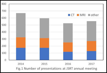
Here, we will introduce the number of presentations at the JSRT annual meeting before COVID-19 (Fig. 1). JSRT is the world’s third largest radiological conference held every April in Yokohama City jointly with JRS and JSMP as JRC. The member presentations to be announced at the JSRT annual meeting are 500 to 600 titles. Research fields at JSRT include general radiography, angiography, CT, MRI, nuclear medicine, ultrasound, education, and more. Among them, CT and MRI account for a half of the total.
3. Recent CT technology
In recent years, CT equipment and CT inspection technology have made remarkable progress. It has never stopped evolving since the development of the EMI scanner by Hounsfield in 1973.
(1). Development of helical scanning
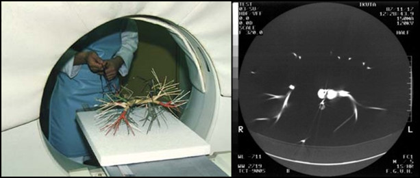
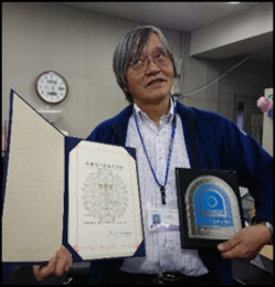
Before the advent of helical scanning, a CT machine was just a machine for tomography. Scanning is performed for each target cross section, and diagnosing human organs requires multiple scans to be performed over time while changing the scanning position. In fact, a lung CT scan required 30 to 40 scans with a slice thickness of 10 mm. This made it impossible to make a correct diagnosis when the respiratory arrest phase was not constant. Therefore, helical scanning using a continuously rotating CT scanner was developed. In helical scanning, the human body moves at a constant speed through a CT device that rotates continuously. This makes it possible to collect three-dimensional data in a single scan. Fig. 2 shows the world’s first helical scan experiment that I conducted in 1987. In the experiment, continuous rotation scans were performed while the lung model was pulled by hand. After that, we created an operating table to move the human body and conducted a basic experiment. This device has been registered as a Future Technology Heritage of the Science and Technology Museum of Japan (Fig. 3).
The clinical advantages of helical scanning include reduced scan time and improved image continuity. Due to the reduction in scan time, lung CT examinations can now scan the entire lung with a single respiratory hold. In addition, it has become possible to reduce the amount of contrast medium used and enhance the contrast enhancement effect. Three-dimensional image diagnosis has become possible by improving the continuity of images.
(2). Development of multi-slice CT
The development of helical scanning changed CT technology from a single scan plane to a stereoscopic scan. Then, multi-slice CT with multiple rows of detectors was developed to realize faster scanning. With the advent of multi-slice CT, it has become possible to perform faster and more detailed scans than ever before.
(3). Development of 3D display
The development of helical scanning and multi-slice CT has greatly changed diagnostic imaging. Along with this, 3D display technology has also evolved. Computers have made it possible to easily display three-dimensional structures that radiologists had previously understood empirically. There are many techniques for three-dimensional image diagnosis, such as MPR (multi-planar reconstruction) diagnosis, MIP (maximum intensity projection), MinIP (minimum intensity projection), and RaySUM (ray-summation).
(4). Development of contrast medium injection techniques
With the development of helical scanning and multi-slice CT, contrast medium injection techniques have changed significantly. These newer CT machines allow for shorter scanning times, but it is difficult to inject contrast media accordingly. Optimal contrast injection techniques allow less contrast to be used. And a more effective contrast enhancement effect can be obtained.
(5). Development of image reconstruction method
The biggest problem with CT is X-ray exposure. In digital images, the higher the X-ray intensity, the smaller the image noise and the more detailed diagnosis becomes possible. For this reason, there was a time when patients’ exposure to radiation increased with the advent of CT. However, as the image reconstruction method has changed from the conventional FBP method to the IR method and DLR, it has become possible to obtain images sufficient for diagnosis with a small X-ray output.
(6). Development of ultra-high-Resolution CT
A high-resolution CT device is required for detailed diagnosis. Factors that affect CT spatial resolution include detector size, focal spot size, and detector data acquisition rate. Canon Medical System has realized ultra-high-resolution CT with a 0.25mm detector.
(7). Development of area-detector CT (ADCT) and Four-dimensional CT (4DCT)
CT diagnostic technology has evolved from 3D to 4D. In helical scanning, the start and end times of scanning are different in order to obtain volume data for spiral rotation. With the area detector CT (ADCT), it is possible to acquire volume data with a single non-helical scan. Four-dimensional CT is possible by performing continuous rotation with this CT device. Fig. 4 is Canon Aquilion ONE with 320 rows x 0.5mm detector. One shot 3D scanning is also possible with this CT, which allows volume data to be obtained in a single scan. This is useful in emergency medicine.
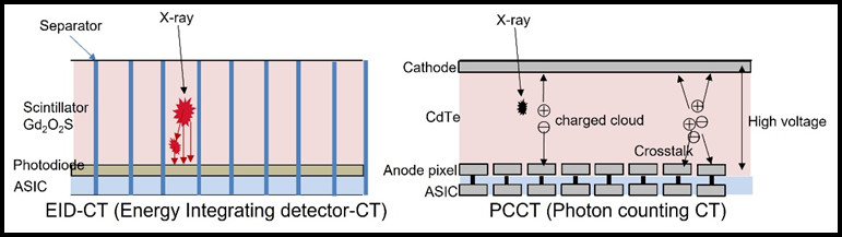
8. Development of Dual Energy CT and Photon counting CT
The latest CT technologies include Dual Energy CT and Photon Counting CT(Fig. 5). With conventional CT, it was not possible to distinguish different materials if they had the same X-ray absorption behavior. Dual Energy CT (DECT) and Photon Counting CT (PCCT) distinguish and detect the X-ray energy transmitted through the object. Then tissue discrimination of iodine, fat, calcium, etc. becomes possible.
4. Recommendations for AFOMP Members
CT is a very important diagnostic device in modern medicine. Treatment begins with a CT diagnosis. Academic activities related to CT will contribute to global medical care. It is the members of Medical Physics who can carry out these activities. We believe that the widespread development of CT and MRI research in the field of medical physics will lead to the development of medical care in your country. Let’s start CT and MRI research together.