
Sourced by A/Prof Vanessa Panettieri, Editor (Educational), AFOMP Pulse
Welcome back to our “Featured Papers” section!
For this September edition, our team has once again explored the AFOMP journals : Physical and Engineering Sciences in Medicine, Journal of Medical Physics, and Radiological Physics and Technology, and selected a range of topics, including some valuable contributions from authors across our beautiful region.
Our first topic focuses on proton therapy, an increasingly important modality across AFOMP countries. This emerging technique continues to evolve, with ongoing developments in precision dosimetry, advanced modelling, and clinical optimisation. Our selection brings together three complementary studies highlighting different aspects of the field: from understanding and accurately measuring proton dose, to using simulation to characterise beam behaviour, and ultimately to optimising the allocation of limited proton-therapy resources to patients.
The second topic is one of my all-time favourites: motion management in radiotherapy. The first paper explores the use of adaptive techniques for patients undergoing SBRT for pancreatic cancer. As we know, the pancreas presents a particularly challenging anatomical site for treatment and can greatly benefit from a well-established motion-management strategy. The second paper examines the use of ultrasound for intrafraction organ-motion tracking during abdominal SBRT.
Finally, we are pleased to reintroduce our “How-to?” section, featuring practical tips on making use of expired EBT-XD radiochromic film, as well as exploring novel applications of the Machine Performance Check tool for linear accelerators.
Happy reading! As always, we welcome your suggestions for topics to explore in future issues and please let us know if you would like to see your next paper featured in this section..
With contributions kindly provided by Sadia Aftab, Medical Physicists, Peter MacCallum Cancer Centre (Australia)
1) Focus on proton therapy. From beam physics to smarter clinical delivery
DOI: 10.1007/s13246-026-01759-7
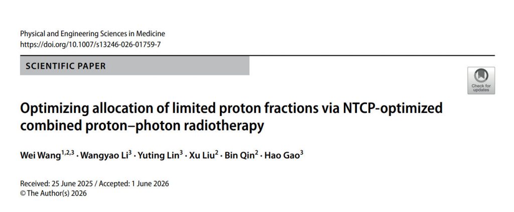
This study by Wang W et al., published in the Physical and Engineering Sciences in Medicine, focused on developing a more efficient way to use limited and expensive proton radiotherapy resources by developing an NTCP-optimised combined proton–photon radiotherapy (NTCP-CPPT) approach. The authors aimed at determining the minimum number of proton fractions needed for effective head-and-neck cancer treatment while still achieving a clinically meaningful reduction in normal-tissue complications, particularly xerostomia and dysphagia.
The main findings showed that incorporating NTCP directly into treatment planning improved organ-at-risk sparing without compromising target coverage, and that the proposed NTCP-CPPT method reduced the average number of proton fractions from 18 to only 3 compared with conventional combined proton–photon planning, while maintaining more than a 20% reduction in summed NTCP relative to conventional IMRT.
The benefits of this work are potentially substantial, including better utilisation of scarce proton therapy slots, lower treatment costs, increased patient access to proton therapy, and reduced predicted radiation-induced toxicities. However, the study also has important limitations: it included only 12 retrospective head-and-neck cases, used 16-field IMRT rather than clinical VMAT, applied different uncertainty-management strategies for IMRT and IMPT, and relied on NTCP models that may not be fully validated across different institutions and patient populations. In addition, the biological effects of different proton and photon fractionation schedules were not incorporated into the optimisation. Overall, this work suggests that NTCP-guided planning may provide a clinically promising and cost-effective strategy for allocating limited proton therapy resources while preserving meaningful normal-tissue sparing.
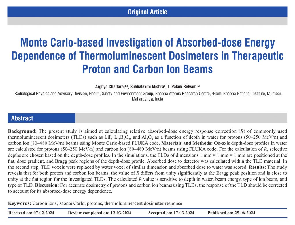
Moving on the Monte Carlo world, the purpose of this work by Chattaraj A et al., published in the Journal of Medical Physics, was to evaluate how accurately commonly used thermoluminescent dosimeters (TLDs) measure absorbed dose in therapeutic proton and carbon-ion beams and to quantify the corrections required because their response is not perfectly water-equivalent. The authors investigated the absorbed-dose energy dependence of three TLD materials—LiF, Li₂B₄O₇, and Al₂O₃—using the FLUKA Monte Carlo simulation code. They calculated the relative absorbed-dose energy response correction factor (R) as a function of depth in water for monoenergetic proton beams (50–250 MeV/n) and carbon-ion beams (80–480 MeV/n), with particular attention to the flat region, dose-gradient region, and Bragg peak of the depth-dose profile.
The results showed that LiF and Li₂B₄O₇ behaved approximately like a water-equivalent material in the flat region, with R values generally within ±5% of unity, whereas Al₂O₃ exhibited larger deviations of about 6–10%. At the Bragg peak, all detectors showed significant energy dependence, with R values varying substantially according to beam energy, ion type, detector material, and detector thickness; overall the largest deviations were observed for Al₂O₃. The study further demonstrated that, in the flat region, detector response is mainly governed by differences in electronic stopping power between the detector material and water. Near the Bragg peak, however, low-energy ions may stop within the detector volume, causing the stopping-power approximation to break down.
Additional simulations with thinner Al₂O₃ detectors showed that reducing detector thickness improves water equivalence, particularly for lower-energy beams.
The main outcome of this work is the provision of beam-specific and depth-specific correction factors for LiF, Li₂B₄O₇, and Al₂O₃ TLDs used in proton and carbon-ion therapy. These findings can improve the accuracy of particle-therapy dosimetry, quality assurance procedures, and detector selection, while also highlighting the importance of considering detector thickness and beam energy in high-LET regions.
Overall, the authors concluded that TLDs cannot be assumed to be universally water-equivalent in proton and carbon-ion therapy and that accurate particle-therapy dosimetry requires appropriate correction factors, particularly near the Bragg peak. The study provides a useful Monte Carlo benchmark for particle-therapy quality assurance and detector characterization, although its direct clinical applicability is limited by the use of idealised monoenergetic beams, a homogeneous water phantom, and the absence of experimental validation and intrinsic thermoluminescence efficiency measurements.
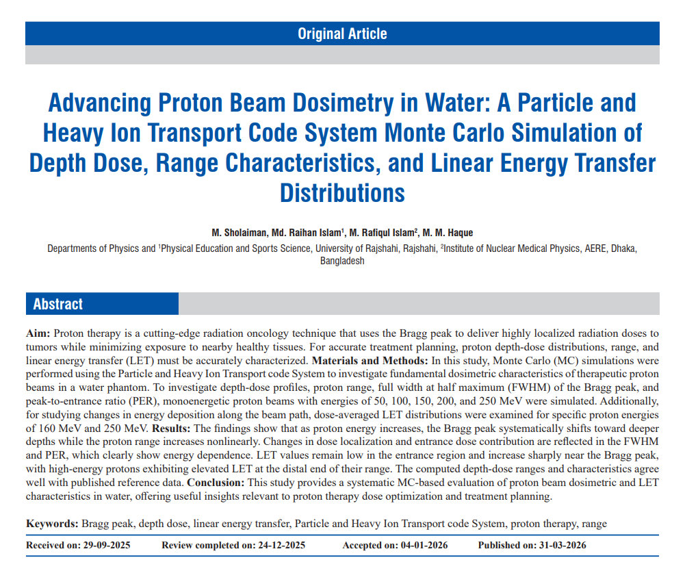
Continuing on the topic of protons this study by Sholaiman M et al., published in the Journal of Medical Physics, investigated proton beam dosimetry using the PHITS Monte Carlo simulation code to evaluate depth-dose distributions, proton range, Bragg peak characteristics, proton fluence, and linear energy transfer (LET) in a water phantom for proton energies between 50 and 250 MeV. The simulations showed that increasing proton energy shifted the Bragg peak to greater depths and increased proton range, while LET remained low in the entrance region and increased sharply near the Bragg peak. The calculated proton ranges agreed closely with established ICRU and NIST reference data, with differences of approximately 0.6–1.6%, confirming the accuracy of the simulation model. The study demonstrated that PHITS can reliably reproduce clinically relevant proton dosimetric parameters, including full width at half maximum (FWHM) and peak-to-entrance ratio (PER), thereby improving understanding of proton beam behaviour and dose localization.
These findings may support treatment planning system commissioning, quality assurance, dose verification, and the development of LET-guided and biologically optimized proton therapy. In addition, the validated simulation framework provides a useful foundation for future investigations involving patient-specific geometries, relative biological effectiveness (RBE), adaptive proton therapy, and secondary particle dosimetry, with the potential to improve treatment accuracy, tumour control, and normal tissue sparing.
However, the study has several limitations. The simulations were performed in a homogeneous water phantom using ideal monoenergetic proton pencil beams rather than patient-specific CT-based geometries and clinical beam delivery conditions. The analysis focused on physical dose and LET, with LET evaluated only for selected energies, and did not include RBE modelling or biological response assessment. Furthermore, the results were validated against published reference data rather than independent experimental measurements, and secondary neutron or out-of-field doses were not assessed. Therefore, further studies incorporating heterogeneous patient anatomies, clinical treatment plans, biological modelling, and experimental validation will be useful before direct clinical application.
2) Focus on: motion management
DOI: 10.1007/s13246-026-01762-y
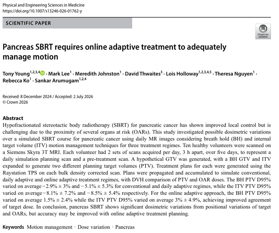
This interesting work by Young T et al., published in Physical and Engineering Sciences in Medicine, examines the challenges of pancreatic stereotactic body radiotherapy (SBRT), particularly the substantial motion of the pancreas caused by respiration, digestion, and normal abdominal organ movement. The study aimed at quantifying dosimetric variations during a simulated pancreatic SBRT course and to compare two motion-management strategies: exhale breath-hold (BH) and free-breathing internal target volume (ITV). It also evaluated three treatment approaches: conventional, daily adaptive, and online adaptive treatment regimes.
Ten healthy volunteers underwent repeated abdominal MRI scans over five days. A hypothetical pancreatic tumour volume was created, and treatment plans were generated for both BH and ITV techniques using RayStation. Dose–volume histogram metrics were used to compare target coverage and organ-at-risk (OAR) doses across all treatment scenarios.
The study found substantial anatomical and dosimetric variations with both BH and ITV techniques. Conventional and daily adaptive approaches showed reduced target coverage and variable OAR doses, particularly for the duodenum. In contrast, online adaptive re-planning maintained target coverage more consistently and generally reduced doses to nearby OARs. The authors concluded that pancreatic SBRT is highly sensitive to day-to-day anatomical changes and that online adaptive treatment provides the most accurate dose delivery.
Important limitations include the use of healthy volunteers rather than patients with pancreatic cancer, so tumour-related and treatment-related anatomical changes were not represented, the small sample size, and the lack of assessment of breath-hold reproducibility and strict control of food and fluid intake.
Overall, this study provides quantitative evidence comparing breath-hold and ITV techniques under conventional, daily adaptive, and online adaptive treatment workflows, showing that online adaptive replanning provides the most reliable target coverage and better protection of nearby organs at risk. For radiation oncologists, medical physicists, and trainees, the study clarifies the limitations of single-plan and same-day replanning approaches and supports the rationale for MRI-guided online adaptive SBRT. It also highlights the importance of motion management, imaging frequency, and patient preparation in abdominal radiotherapy, making the findings directly relevant to treatment planning, quality assurance, and future research design
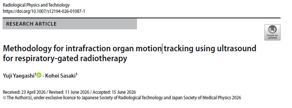
DOI: 10.1007/s12194-026-01087-1
This interesting work by Yaegashi Y et al., published in Radiological Physics and Technology. Authors aimed to develop and demonstrate a non-invasive ultrasound-based method for tracking intrafraction organ motion during respiratory-gated radiotherapy (SBRT) for non-small cell lung cancer. The authors have proposed a system that automatically detects liver edges using the Shi–Tomasi method and tracks respiratory motion in real time using the Lucas–Kanade optical flow technique, with irradiation timing determined by predefined displacement and image-matching thresholds. Phantom experiments showed successful tracking at respiratory rates of 12 and 15 breaths per minute, while tracking was not consistently maintained at 20 breaths per minute. In addition, tests in five healthy volunteers confirmed that liver motion could be successfully tracked during natural breathing, supporting the feasibility of the method in humans. The main benefits of this approach are that it is non-invasive, avoids implanted fiducial markers and additional X-ray exposure, provides real-time visualization of internal organ motion, and may offer more accurate respiratory gating than external abdominal marker systems.
However, the study also identified important limitations, including the challenge of reliably fixing the ultrasound probe on the abdomen during natural breathing, the fact that the method remains a surrogate for actual tumor position and therefore still requires confirmation with EPID or X-ray fluoroscopy before treatment, and the need for further research to address liver deformation and to evaluate applicability to organs other than the lungs.
3) Focus on: How to?
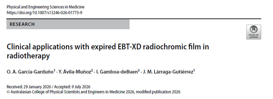
DOI: 10.1007/s13246-026-01773-9
This very interesting study by García-Garduño A et al., published in Physical and Engineering Sciences in Medicine, investigated whether expired EBT-XD radiochromic films can still be used for selected radiotherapy dosimetry applications. The work addresses an important clinical issue, as radiotherapy departments often accumulate expired film stock due to budget constraints, and the reuse of these films could provide both economic and environmental benefits.
The authors compared two expired EBT-XD film batches with a non-expired batch using a 6 MV photon beam. They evaluated dose–response behaviour, small-field output factors, and patient-specific quality assurance (PSQA) for volumetric-modulated arc therapy (VMAT) and stereotactic radiosurgery (SRS). The results showed that expired films retain a measurable dose-dependent response but exhibit reduced sensitivity, particularly at low doses. After generating new calibration curves, the expired films demonstrated acceptable agreement with non-expired films for small-field output-factor measurements and high-dose SRS PSQA, with gamma pass rates generally exceeding 98%. In contrast, performance was less reliable for low-dose VMAT QA, where increased uncertainty and reduced sensitivity affected agreement with the planned dose distributions.
The study concludes that properly stored and recalibrated expired EBT-XD films may still be useful as relative dosimeters for selected moderate to high-dose radiotherapy measurements when used within the recommended dose range. However, caution is required for low-dose measurements and for films that have been expired for many years.
A major strength of this work is that it provides clinically relevant evidence supporting the potential reuse of expired EBT-XD films in selected applications, offering possible cost savings, reduced material waste, and practical guidance for small-field dosimetry and high-dose SRS patient-specific QA.
The study also has several limitations. The low-dose VMAT analysis was based on only a small number of representative treatment plans and expired film samples, limiting the statistical robustness and generalisability of the findings. In addition, only three EBT-XD batches stored under controlled conditions were evaluated using a single 6 MV photon beam and one linear accelerator platform; therefore, the results cannot be directly extrapolated to other beam qualities, storage conditions, or dosimetric applications such as brachytherapy or leakage measurements. Finally, reduced sensitivity in older expired films may artificially improve gamma pass rates, particularly in low-dose regions, introducing additional uncertainty in the interpretation of PSQA results.
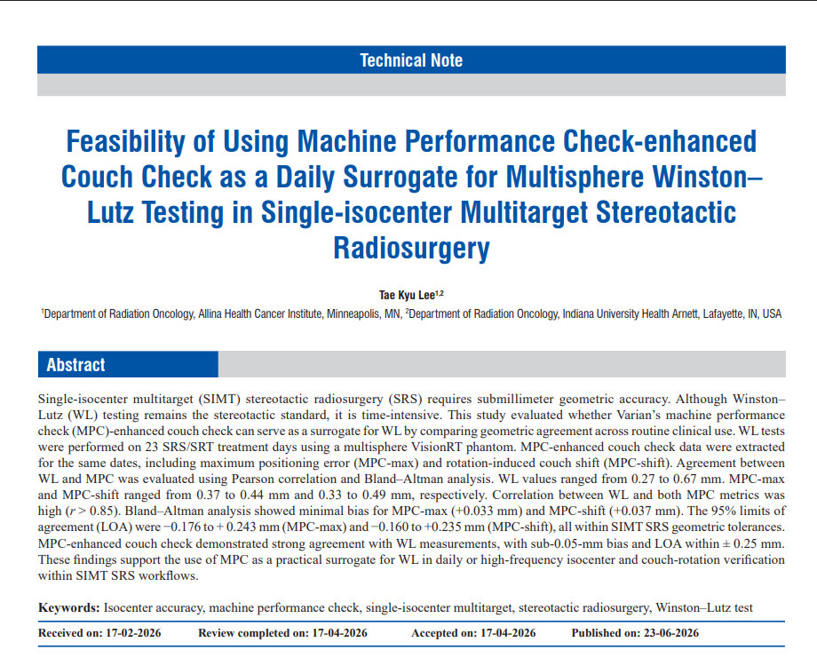
This feasibility study by Lee T.K published in the Journal of Medical Physics, was conducted to determine whether Varian’s Machine Performance Check enhanced couch check (MPC-EC) could be used as a practical daily alternative to the multisphere Winston–Lutz (WL) test for single-isocenter multitarget stereotactic radiosurgery (SIMT SRS), where submillimeter geometric accuracy is critical. The need for this work arose because conventional WL testing, although considered the clinical gold standard for stereotactic isocenter verification, is labour-intensive and difficult to perform every treatment day in high-volume clinics. The authors compared paired WL and MPC measurements acquired on 23 SRS/SRT treatment days and found strong agreement between the two methods, with correlation coefficients greater than 0.86, mean biases below 0.04 mm, and 95% limits of agreement within approximately ±0.25 mm, all well within accepted SIMT SRS geometric tolerances. These findings indicate that MPC can reliably track daily isocenter stability and couch-rotation accuracy, even on days with the largest observed WL deviations. The main benefit of the approach is improved clinical efficiency, MPC can be completed in less than five minutes and is suitable for routine daily use, potentially reducing workload while maintaining stereotactic-level precision. However, the study was limited by its relatively small sample size, single-institution design, and evaluation over only 23 treatment sessions, and it did not replace the value of periodic WL testing for commissioning, baseline establishment, or detailed off-axis verification. Overall, the outcome of the work supports the feasibility of using MPC-enhanced couch check as a reliable surrogate for daily or high-frequency WL verification in SIMT SRS workflows, allowing clinics to streamline quality assurance without compromising geometric accuracy.